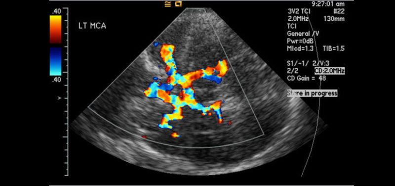
Furthermore, approximately 10–20% of patients have inadequate transtemporal acoustic windows.

It also has a long learning curve to acquire the three-dimensional understanding of cerebrovascular anatomy necessary for competency. The technique is however highly operator dependent, which can significantly limit its utility. It is relatively inexpensive, repeatable, and its portability offers increased convenience over other imaging methods, allowing continuous bedside monitoring of CBF-V, which is particularly useful in the intensive care setting. TCD allows dynamic monitoring of cerebral blood flow velocity (CBF-V) and vessel pulsatility over extended time periods with a high temporal resolution. Transcranial Doppler (TCD), first described in 1982, is a noninvasive ultrasound (US) study that involves the use of a low-frequency (≤2 MHz) transducer probe to insonate the basal cerebral arteries through relatively thin bone windows. TCD is also used in brain stem death, head injury, raised intracranial pressure (ICP), intraoperative monitoring, cerebral microembolism, and autoregulatory testing. Current applications of TCD include vasospasm in sickle cell disease, subarachnoid haemorrhage (SAH), and intra- and extracranial arterial stenosis and occlusion. However, the performance of TCD is highly operator dependent and can be difficult, with approximately 10–20% of patients having inadequate transtemporal acoustic windows.

It is relatively inexpensive, repeatable, and portable. TCD allows dynamic monitoring of CBF-V and vessel pulsatility, with a high temporal resolution. It involves use of low-frequency (≤2 MHz) US waves to insonate the basal cerebral arteries through relatively thin bone windows. Transcranial Doppler (TCD) is a noninvasive ultrasound (US) study used to measure cerebral blood flow velocity (CBF-V) in the major intracranial arteries.


 0 kommentar(er)
